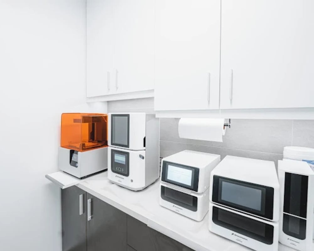
In-House Dental Laboratory
At Centre Dentaire St-Onge, we are proud to have our own on-site dental laboratory. This allows us to create dental prosthetics directly in-house.
Advantages of Our Laboratory
-
Speed & Efficiency
Thanks to our on-site laboratory, we can significantly reduce wait times for the fabrication and adjustment of dental prosthetics. Our patients benefit from faster restorations without compromising on quality.
-
Technology
We use dental implant and milling technology like the Straumann P30. This helps us fabricate highly precise prosthetics and ensure an optimal fit with enhanced durability.
-
Materials
These materials help ensure that our restorations are not only functional but also aesthetically pleasing.
-
Quality Control
With our in-house laboratory, we maintain full control over every step of the fabrication process. Our dental technicians work closely with our dentists to help ensure each prosthetic meets high standards of quality and precision.
- Personalization & Flexibility
Guided Dental Surgery
SimPlant® is implant treatment planning software that provides the dentist with three-dimensional (3D) data on the width and density of the jawbone, the exact location of the roots of the teeth adjacent to the surgical site, and the position of the nerves.
So what does this mean? The practitioner can plan the ideal placement of the implant, determine the expected results, and even show the patient what their smile could look like.
SimPlant allows for highly personalized treatment. It is fast and efficient, and helps reduce unexpected outcomes, complications, and long recovery times.
Guided Surgeries
Guided surgery in dental implantology is an approach that uses digital technology to help plan and precisely perform the placement of dental implants. Here is a detailed description of this method.
-
Learn More
1. Precise Virtual Planning:
Guided surgery begins with the acquisition of radiographic images and the digital scanning of the patient’s jaw using CBCT (Cone Beam Computed Tomography) scanners and other 3D imaging technologies. This data makes it possible to create a detailed digital representation of the jaw and surrounding structures.
2. Implant Position Planning:
Using computer-aided planning software (CAD), the dental surgeon can visualize the patient’s jaw in 3D and accurately determine the ideal position for each implant. This includes consideration of bone density, important anatomical structures, and prosthetic treatment plans.
3. Fabrication of Custom Surgical Guides:
Based on the virtual planning, custom surgical guides are created. These guides are 3D-printed and used during the procedure to accurately guide the position and angle of dental implant placement.
4. Precise and Minimally Invasive Surgery:
During the procedure, the surgical guide is placed in the patient’s mouth, providing precise guidance for each incision and implant placement. This helps reduce the risk of surgical errors, minimizes trauma to surrounding tissues, and allows for more predictable results.
5. Benefits for Patients:
Guided surgery in dental implantology offers several benefits for patients, including reduced surgical time, decreased post-operative discomfort, faster recovery, and improved aesthetic outcomes.
6. Improved Communication & Interdisciplinary Collaboration:
This method also facilitates communication between practitioners, ensuring smooth integration between the surgical phase and the prosthetic restoration phase.
Solea
The Solea system is a major advancement in laser dentistry, offering several significant advantages compared to traditional techniques.
-
Learn More
1. High Precision:
Solea uses laser technology that allows for dental procedures with a very high level of precision. This enables dentists to target affected areas while preserving the surrounding healthy tooth structure.
2. Virtually Pain-Free Procedures:
Unlike traditional drills, Solea is virtually pain-free. The laser simultaneously seals blood vessels, minimizing bleeding and reducing the need for local anesthesia in many cases.
3. Reduced Trauma:
By eliminating the vibrations and noise associated with conventional drills, Solea helps reduce stress and anxiety for patients. This makes dental visits more comfortable and less intimidating.
4. Faster Healing:
By preserving more healthy tissue and helping reduce the risk of complications, Solea supports faster healing after dental procedures. Patients recover more quickly and can return to their normal activities sooner.
5. Versatility:
Solea can be used for a wide range of dental procedures, including cavity treatment, tooth preparation, repair of existing restorations, and even gum procedures. This makes it a versatile tool for dentists looking to provide quality dental care.
Planmeca Viso G5
Digital radiology in dental care uses technology to capture and process radiographic images digitally, offering several advantages over conventional radiology. Here is a detailed description of this technology and its main advantages.
-
Learn More
1. Digital Image Capture:
Instead of using traditional radiographic film, digital radiology uses sensitive electronic sensors to record X-rays passing through dental and bone tissues.
2. Computer Processing:
The captured signals are instantly converted into digital images and displayed on a computer screen. This allows for immediate viewing and easy manipulation of radiographic images.
3. Sensor Technologies:
Sensors can be intraoral (placed directly in the patient’s mouth) or extraoral (used for panoramic images or CT scans).
4. Digital Storage & Archiving:
Digital images can be easily stored in computerized systems (PACS – Picture Archiving and Communication System) and accessed at any time, facilitating long-term archiving and access to patients’ radiographic records.
5. Advantages
- Reduced radiation exposure
- Improved image quality
- Efficiency and speed
- Cleaner working environment
- Improved communication and collaboration
- Increased patient comfort
CEREC
The CEREC system (Chairside Economical Restoration of Esthetic Ceramics) is a technology used in restorative dentistry that offers significant advantages for both dentists and patients. Here is a detailed description of its main features.
-
Learn More
1. Computer-Aided Design (CAD):
CEREC uses CAD technology to create dental restorations such as crowns, inlays, onlays, and veneers directly at the dental clinic. This eliminates the need for multiple visits and uncomfortable temporary restorations.
2. 3D Optical Imaging:
The process begins with the precise digital capture of the dental impression using an intraoral camera. This 3D optical imaging replaces traditional techniques that are often uncomfortable and less accurate.
3. Virtual Design & On-Site Fabrication:
Once the digital impression is obtained, the CEREC software allows the dentist to design the restoration directly on a computer screen. The virtual model is then sent to a milling unit, which shapes the restoration from high-quality ceramic in a short amount of time.
4. Aesthetic & Durable Restorations:
The ceramic materials used by CEREC are known for their aesthetics and durability. They are designed to match the color and translucency of natural teeth, providing a natural-looking result.
5. Comfort & Convenience for Patients:
With CEREC, patients receive high-quality, custom dental restorations in a single visit. This reduces time spent at the dental clinic, minimizes discomfort associated with longer procedures, and eliminates the need for temporary restorations.
6. Reduced Errors and Adjustments:
By using digital technology, CEREC significantly reduces the risk of errors associated with manual impressions and the adjustments often required with traditional restorations.
Digital X-Rays
Digital radiology in dentistry uses modern technology to capture and process radiographic images digitally, offering several significant advantages over conventional radiology. Here is a detailed description of this technology and its main advantages.
-
Learn More
1. Digital Image Capture:
Instead of using traditional radiographic film, digital radiology uses sensitive electronic sensors to record X-rays passing through dental and bone tissues.
2. Computer Processing:
The captured signals are instantly converted into digital images and displayed on a computer screen. This allows for immediate viewing and easy manipulation of radiographic images.
3. Sensor Technologies:
Sensors can be intraoral (placed directly in the patient’s mouth) or extraoral (used for panoramic images or CT scans).
4. Digital Storage & Archiving:
Digital images can be easily stored in computerized systems (PACS – Picture Archiving and Communication System) and accessed at any time, facilitating long-term archiving and access to patients’ radiographic records.
5. Advantages:
- Reduced radiation exposure
- Improved image quality
- Efficiency and speed
- Cleaner working environment
- Improved communication and collaboration
- Increased patient comfort
CariVu
The CariVu system by Dexis is a technology designed to improve the detection of dental caries in a non-invasive and precise way. Here are its main features and advantages.
-
Learn More
1. Transillumination Imaging:
CariVu uses transillumination, a technique that projects a specific light through the tooth. This allows dentists to clearly visualize the internal structures of the tooth without ionizing radiation, unlike traditional X-rays.
2. Early & Accurate Detection of Cavities:
By illuminating the tooth from within, CariVu reveals cavities, even those that are very small or located beneath the surface of the enamel. This helps dentists detect cavities at an early stage, allowing for more conservative and less invasive treatment.
3. Ease of Use:
The device is easy to handle and integrate into daily dental practice. Images are instantly displayed on a screen, allowing patients to see the affected areas and better understand their dental health.
4. Reduced Radiation Exposure:
Since it does not use X-rays, CariVu is especially beneficial for patients who prefer to avoid radiation exposure, such as pregnant women and children.
5. Complementary to Other Diagnostic Methods:
Although CariVu is very effective for detecting cavities, it can also be used alongside traditional X-rays to provide a more complete diagnostic assessment.
PRGF-ENDORET
PRGF (Plasma Rich in Growth Factors) and PRF (Platelet-Rich Fibrin) treatments are regenerative medicine techniques used to promote the healing and regeneration of both soft and hard tissues. Here is a detailed description of each of these treatments.
-
Learn More
PRGF (Plasma Rich in Growth Factors)
1. Preparation Process:
PRGF is obtained from the patient’s own blood. A small blood sample is drawn, usually using a special syringe, and then centrifuged to separate the different blood components.
2. Concentration of Growth Factors:
Centrifugation concentrates platelets rich in growth factors into a small volume of plasma. Growth factors are natural proteins that play a crucial role in tissue regeneration and healing.
3. Clinical Application:
Once prepared, PRGF is applied directly to the treatment area, for example during dental implant placement, bone grafting, or periodontal surgery. It can be used in gel form or injected, depending on the specific needs of the treatment.
4. Advantages:
- Promotes fast and predictable tissue healing
- Reduces the risk of post-operative complications.
- Improves the quality and quantity of regenerated tissue
- Uses the patient’s own blood, reducing the risk of incompatibility or allergic reaction
PRF (Platelet-Rich Fibrin)
1. Preparation Process:
PRF is also obtained from the patient’s blood, but the centrifugation process is slightly different. It focuses on forming a fibrin clot rich in platelets and growth factors.
2. PRF Structure:
PRF appears as a dense fibrin gel rich in platelets and leukocytes. This gel contains a high concentration of growth factors and cytokines that promote tissue regeneration.
3. Clinical Application:
PRF is mainly used to enhance healing and regeneration of soft tissues, such as closing graft sites, treating periapical lesions, and periodontal regeneration procedures.
4. Advantages:
- Stimulates soft tissue regeneration
- Improves the quality of post-operative healing
- Promotes the formation of new blood vessels and bone growth
Comparison & Choice
PRGF vs PRF:
Although similar in their basic concept (using growth factors from the patient’s blood), PRGF focuses on separating platelet-rich plasma, while PRF is more focused on forming a dense fibrin gel. The choice between the two depends on the specific needs of the treatment and the practitioner’s preference.